Chlamydia: различия между версиями
Материал из Поле цифровой дидактики
| Строка 10: | Строка 10: | ||
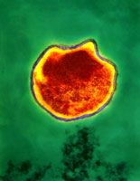
https://microbewiki.kenyon.edu/images/1/11/Chlamydia_200.jpg | https://microbewiki.kenyon.edu/images/1/11/Chlamydia_200.jpg | ||
https://upload.wikimedia.org/wikipedia/commons/a/aa/ChlamydiaTrachomatisEinschlusskörperchen.jpg | |||
Версия 15:16, 23 сентября 2022
| Описание микроба | [[Description::Chlamydia has long plagued humanity as the most commonly contracted STD, caused by Chlamydia trachomatis. With the sequencing of the C. trachomatis genome, our ability of understand, diagnose, and combat the pathogen is greatly increased.]] |
|---|---|
| Верхний таксон | Bacteria; Chlamydiae/Verrucomicrobia group; Chlamydiae; Chlamydiae (class); Chlamydiales; Chlamydiaceae; Chlamydia |
| Species - виды | Chlamydia muridarum, Chlamydia trachomatis, Chlamydia pneumoniae (also known as Chlamydophila pnemoniae) |
| Genome Structure= структура генома | The sequences of both Chlamydia trachomatis and Chlamydia pneumoniae have been determined with the hope that a comparison between the two genomes will significantly enhance the understanding of both pathogens. Identification of genes particular to one or the other species could indicate mutually exclusive biological, virulence, and pathogenesis capabilities, while genes the two have in common will help researchers better understand the metabolic capabilities necessary for living in a human host. |
| Cell Structure - клеточная структура | Both Chlamydia trachomatis and Chlamydia pneumoniae are Gram-negative (or at least are classified as such, they are difficult to stain, but are more closely related to Gram-negative bacteria), aerobic, intracellular pathogens. They are typically coccoid or rod-shaped and require growing cells to remain viable. Chlamydia cannot synthesize its own ATP, and can also not be grown on an artificial medium, and consequently was once thought to be a virus. The unique cell wall of Chlamydia trachomatis is thought to be one of its virulence factors, as it inhibits phagolysosome fusion in phagocytes. The cell wall contains an outer lipopolysaccharide membrane but lacks peptidoglycan. It instead contains cysteine-rich proteins that are likely the functional equivalent of peptidoglycan. This unique cell wall structure, allows for intracellular division and extracellular survival. |
| Ecology= экология обитания | Chlamydia has a very unique life-cycle, in which in alternates between a non-replicating, infectious elementary body, and a replicating, non-infectious reticulate body. The elementary body is the dispersal form of the pathogen, and is analogous to the spore. The bacterium induces its own endocytosis upon contact with potential host cells. Once inside a cell the elementary body germinates as the result of interaction with glycogen, and converts to its vegetative, reticulate form. The reticulate form divides every 2-3 hours, and has an incubation period of about 7-21 days in its host. After division, the pathogen reverts back to its elementary form and is released by the cell through exocytosis. They are extremely temperature sensitive, and must be refrigerated at 4 degrees celsius as soon as a sample is obtained. |
https://microbewiki.kenyon.edu/index.php/Chlamydia


